Fichier:Staphylococcus aureus VISA 2.jpg

Fichier d’origine (1 420 × 1 091 pixels, taille du fichier : 259 kio, type MIME : image/jpeg)
Il peut être utilisé par d'autres projets. Sa description sur sa page de description est affichée ci-dessous. |
| Description |
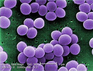
English: Under a very high magnification of 20,000x, this scanning electron micrograph (SEM) shows a strain of Staphylococcus aureus bacteria taken from a vancomycin intermediate resistant culture (VISA). Under SEM, one can not tell the difference between bacteria that are susceptible, or multidrug resistant, but with transmission electron microscopy (TEM), VISA isolates exhibit a thickening in the cell wall that may attribute to their reduced susceptibility to vancomycin . See PHIL 11156 for a black and white version of this image. VISA and VRSA are specific types of antimicrobial-resistant staph bacteria. While most staph bacteria are susceptible to the antimicrobial agent vancomycin some have developed resistance. VISA and VRSA cannot be successfully treated with vancomycin because these organisms are no longer susceptibile to vancomycin. However, to date, all VISA and VRSA isolates have been susceptible to other Food and Drug Administration (FDA) approved drugs. How do VISA and VRSA get their names? Staph bacteria are classified as VISA or VRSA based on laboratory tests. Laboratories perform tests to determine if staph bacteria are resistant to antimicrobial agents that might be used for treatment of infections. For vancomycin and other antimicrobial agents, laboratories determine how much of the agent it requires to inhibit the growth of the organism in a test tube. The result of the test is usually expressed as a minimum inhibitory concentration (MIC) or the minimum amount of antimicrobial agent that inhibits bacterial growth in the test tube. Therefore, staph bacteria are classified as VISA if the MIC for vancomycin is 4-8µg/ml, and classified as VRSA if the vancomycin MIC is >16µg/ml. |
||
| Date | |||
| Source |
|
||
| Auteur |
Content Providers(s): CDC/ Matthew J. Arduino, DRPH |
||
| Autorisation (Réutilisation de ce fichier) |
PD-USGov-HHS-CDC English: None - This image is in the public domain and thus free of any copyright restrictions. As a matter of courtesy we request that the content provider be credited and notified in any public or private usage of this image. |
Cette image est l’œuvre des
Centers for Disease Control and Prevention , division du Département de la Santé et des Services Sociaux des États-Unis, réalisée par un employé dans le cadre de ses activités professionnelles. En tant qu'œuvre du gouvernement fédéral des États-Unis d'Amérique, cette image est placée dans le domaine public.
|
Historique du fichier
Cliquer sur une date et heure pour voir le fichier tel qu'il était à ce moment-là.
| Date et heure | Vignette | Dimensions | Utilisateur | Commentaire | |
|---|---|---|---|---|---|
| actuel | 3 août 2009 à 22:24 | | 1 420 × 1 091 (259 kio) | wikimediacommons>Raeky | {{Information |Description={{en|1='''Under a very high magnification of 20,000x, this scanning electron micrograph (SEM) shows a strain of Staphylococcus aureus bacteria taken from a vancomycin intermediate resistant culture (VISA).'''<p> Under SEM, one |
Utilisation du fichier
Les 2 pages suivantes utilisent ce fichier :