Fichier:Pyogenic granuloma on a finger-1.jpg

Taille de cet aperçu : 800 × 600 pixels. Autres résolutions : 320 × 240 pixels | 640 × 480 pixels | 1 024 × 768 pixels | 1 280 × 960 pixels | 2 560 × 1 920 pixels | 3 648 × 2 736 pixels.
Fichier d’origine (3 648 × 2 736 pixels, taille du fichier : 3,5 Mio, type MIME : image/jpeg)
Il peut être utilisé par d'autres projets. Sa description sur sa page de description est affichée ci-dessous. |
| Description |
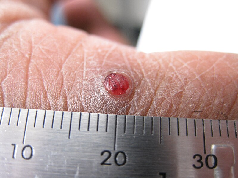
English: A pyogenic granuloma situated on the dorsal surface of an index finger. It is a solitary papule of inflamed vascular granulation tissue. Pyogenic granulomas can grow over a few weeks from a break in the skin - after a cut or deep graze. They generally occur on the fingers, lips, mouth, trunk, and toes. On the palms and soles they have a typical collar of thickened stratum corneum at the base, which is evident here. These lesions are fragile and tend to bleed after minor trauma. They have variable appearances - they may have a smooth surface or may be covered with a crust or exudate; they may appear bright red, dusky red, violaceous, or brown black in color. After treatment to disrupt the growing granulation tissue they can be expected to heal after 1-3 weeks depending on their initial size. The ruler has hash marks at millimeter intervals. |
| Date | |
| Source | Travail personnel |
| Auteur | kilbad (talk) |
Conditions d’utilisation
Kilbad sur Wikipédia anglais, en tant que détenteur des droits d’auteur sur cette œuvre, la publie sous la licence suivante :
Ce fichier est disponible selon les termes de la licence Creative Commons Attribution 3.0 Non transposée.
Attribution: Kilbad sur Wikipédia anglais
- Vous êtes libre :
- de partager – de copier, distribuer et transmettre cette œuvre
- d’adapter – de modifier cette œuvre
- Sous les conditions suivantes :
- paternité – Vous devez donner les informations appropriées concernant l'auteur, fournir un lien vers la licence et indiquer si des modifications ont été faites. Vous pouvez faire cela par tout moyen raisonnable, mais en aucune façon suggérant que l’auteur vous soutient ou approuve l’utilisation que vous en faites.
Journal des téléversements d’origine
Transféré de en.wikipedia à Commons par Snowmanradio utilisant CommonsHelper.
La page de description originale était ici. Tous les noms d'utilisateur qui suivent se rapportent à en.wikipedia.
- 2010-02-24 02:54 Kilbad 3648×2736× (3673609 bytes) {{Information |Description = A solitary [[pyogenic granuloma]] |Source = I (~~~) created this work entirely by myself. |Date = ~~~~~ |Author = ~~~ |other_versions = }}
Historique du fichier
Cliquer sur une date et heure pour voir le fichier tel qu'il était à ce moment-là.
| Date et heure | Vignette | Dimensions | Utilisateur | Commentaire | |
|---|---|---|---|---|---|
| actuel | 25 février 2010 à 15:36 | | 3 648 × 2 736 (3,5 Mio) | wikimediacommons>File Upload Bot (Magnus Manske) | {{BotMoveToCommons|en.wikipedia|year={{subst:CURRENTYEAR}}|month={{subst:CURRENTMONTHNAME}}|day={{subst:CURRENTDAY}}}} {{Information |Description={{en|A en:pyogenic granuloma situated on the dorsal surface of an index finger. It is a solitary [[:en |
Utilisation du fichier
La page suivante utilise ce fichier :