Fichier:Gray491.png
Gray491.png (500 × 438 pixels, taille du fichier : 63 kio, type MIME : image/png)
Il peut être utilisé par d'autres projets. Sa description sur sa page de description est affichée ci-dessous. |
Description
| Description |
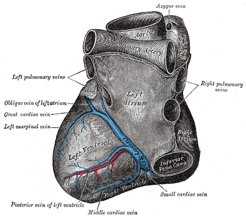
Deutsch: Blick von hinten auf das Herz. Darstellung von Henry Gray. |
||||||||||||||||||||
| Planche | 491 | ||||||||||||||||||||
| Date | avant 1858 | ||||||||||||||||||||
| Source |
|
||||||||||||||||||||
| Auteur |
|
||||||||||||||||||||
.jpg)
Livre
| Henry Gray : Gray's Anatomy (20e édition)
|
|||||||||||||||||||||||
|---|---|---|---|---|---|---|---|---|---|---|---|---|---|---|---|---|---|---|---|---|---|---|---|
| Auteur |
|
-_Title_page.png) | |||||||||||||||||||||
| Correcteur |
Revised by Warren H. Lewis |
||||||||||||||||||||||
| Illustrateur |
|
||||||||||||||||||||||
| Titre | |||||||||||||||||||||||
| Édition |
20 |
||||||||||||||||||||||
| Éditeur de publication | |||||||||||||||||||||||
| Type d'objet |
version, édition ou traduction |
||||||||||||||||||||||
| Liste des pages | list of all the plates | ||||||||||||||||||||||
| Langue |
anglais |
||||||||||||||||||||||
| Date de publication |
1918 |
||||||||||||||||||||||
| Lieu de publication |
Philadelphie / New York |
||||||||||||||||||||||
| Source | Bartleby | ||||||||||||||||||||||

Conditions d’utilisation
Cette image est dans le domaine public car elle a été obtenue en scannant ou en photocopiant l'original qui est lui-même dans le domaine public, ou car elle est tellement similaire à un scan ou à une photocopie qu'aucun nouveau droit d'auteur n'a été créé. L'original est dans le domaine public pour la raison suivante :
Ce modèle est destiné à être utilisé dans des situations où il peut être nécessaire d'expliciter que des améliorations (par ex. luminosité, contraste, égalisation des couleurs, amélioration de la netteté) sont en elles-mêmes insuffisantes pour donner lieu à de nouveaux droits d'auteur. Ce modèle peut être utilisé lorsqu'on ne sait pas si des améliorations ont été apportées ainsi que quand des améliorations sont visibles mais ne génèrent pas de nouveaux droits. Si vous savez qu'un scan n'a pas été amélioré, utilisez {{PD-old}}. Pour plus d'informations, voir Commons:Quand utiliser le bandeau PD-scan.  | ||||
The coronary sinus is a collection of veins joined together to form a large vessel that collects blood from the myocardium of the heart. It is present in humans and other animals. It delivers deoxygenated blood to the Right atrium in conjunction with the superior and inferior vena cava.
The coronary sinus opens into the right atrium, between the inferior vena cava and the atrio-ventricular orifice. It returns the blood from the substance of the heart, and is protected by a semicircular fold of the lining membrane of the auricle, the coronary valve (the valve of Thebesius). The sinus, before entering the auricle, is considerably dilated - nearly to the size of the end of the little finger. Its wall is partly muscular, and at its junction with the great coronary vein is somewhat constricted and furnished with a valve consisting of two unequal segments.(Gray 462)
Location: It is located in the right atrium and runs transversely in the groove between the left atrium and ventricle on the posterior surface of the heart.
The coronary sinus orifice (opening) is just superior to the septal leaflet of the tricuspid valve. The coronary sinus orifice is also known as the ostium of the coronary sinus, and is guarded by the Thebesian valve.
Drainage: It receives blood mainly from the small, middle, great and oblique cardiac veins. It also receives blood from the left marginal vein and the left posterior ventricular vein. The anterior cardiac veins drain directly into the right atrium. (Some small veins drain into any of the four chambers of the heart.)
It drains into the right atrium on the posterior, inferior surface, medial to the inferior vena cava opening.
Historique du fichier
Cliquer sur une date et heure pour voir le fichier tel qu'il était à ce moment-là.
| Date et heure | Vignette | Dimensions | Utilisateur | Commentaire | |
|---|---|---|---|---|---|
| actuel | 23 janvier 2007 à 16:35 | | 500 × 438 (63 kio) | wikimediacommons>Pngbot | optimized with optipng |
Utilisation du fichier
La page suivante utilise ce fichier :